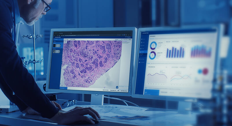

“Glass slides work fine” is often a way of saying that pathologists can’t justify the investment in digital solutions.
DIGITAL PATHOLOGY MANUAL
Speeds up access to samples and improves turnaround time versus manual reviews, especially in complex casesīig data allows pathologists to become more specializedĪllows practices to extend to broader geographiesĭelivers better tools for teaching and training Reduces time retrieving, data matching and organizing The future of digital pathology could eventually encompass enhanced translational research, computer aided diagnosis (CAD) and personalized medicine.Ĭentral storage enables easy access in streamlined workflowĪllows for automation, flex work schedules and remote access
DIGITAL PATHOLOGY SOFTWARE
The rapid progress of whole slide imaging (WSI) technology, along with advances in software applications, LIS / LIMS interfacing, and high-speed networking, have made it possible to fully integrate digital pathology into pathology workflows.ĭigital pathology enables pathologists to engage, evaluate, and collaborate rapidly and remotely, with transparency and consistency, thus improving efficiency and productivity. However, it is in the past decade that pathology has begun to undergo a true digital transformation, moving away from analog into an electronic environment. The concept of telepathology - transmitting microscope images between remote locations - has been around for nearly 50 years.

The history of digital pathology goes back over 100 years, when specialized equipment was first used to capture images from a microscope onto photographic plates. Automated image analysis tools can also be applied to assist in the interpretation and quantification of biomarker expression within tissue sections. Digital slides can be shared over networks using specialized digital pathology software applications. The different tiers of digital pathology | Mike Miller, I.Utilizing high-throughput, automated digital pathology scanners, it is possible to capture an entire glass slide, under bright field or fluorescent conditions, at a magnification comparable to a microscope. A simple microscope camera, whole slide scanner and everything in between.How Pathology Watch managed to incorporate digital pathology in dermatology practices across the US | Dan Lambert, Pathology Watch Microscopic imaging of tissue without a microscope?.Researchers create detailed cell atlas of gut and reveal developmental origins of Crohn’s disease.Breast cancer ‘ecotypes’ defined by cellular & spatial genomics technologies could lead to more personalised treatment.Literature Review: Drug development and toxicologic pathology continue to blaze the trail in digital pathology.Publication Review – Tissues, Not Blood, Are Where Immune Cells Function.Publication Review – The human-in-the-loop: an evaluation of pathologists’ interaction with artificial intelligence in clinical practice.Proscia’s Concentriq Dx Attains CE Mark Under IVDR For Use In Primary Diagnosis.Novel Digital Microscopy System Provides Faster Diagnosis Than Light Microscopy.Groundbreaking digital approach helps cut pathology waiting times.Graph Representation Learning Combines Local and Overall Tissue-Level Information to Predict Disease States.AI and 3D imaging collaborate to Provide Never-Before-Seen View and Understanding of Prostate Cancer Cells.Paige Answers Call to Better Identify Breast Cancer Patients with Low Expression of HER2.New real-time imaging technology could make biopsies & histology a thing of the past.Proscia-Siemens Healthineers: A win-win deal set to shake-up Digital Pathology?.A new tool makes high-resolution imaging data on human tissues easier to understand and use.


 0 kommentar(er)
